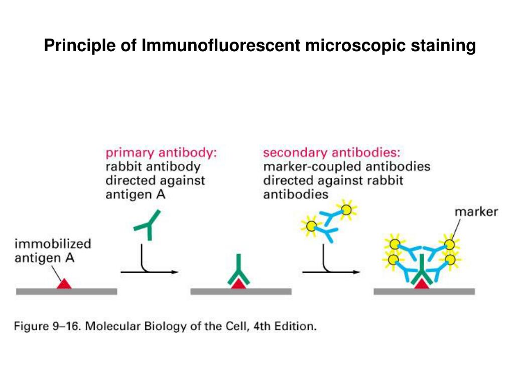
The different approaches to achieve multiplexed IHC and immunofluorescence are described in this study. Alternatives to multiplex immunofluorescence/IHC include epitope-based tissue mass spectrometry and digital spatial profiling (DSP), which require specialized platforms not available to most clinical laboratories.. such as autostainer type.. Immunofluorescence is a type of assay performed on biological samples to detect specific antigens in any biological specimen or sample and vice-versa. The specificity of antibodies to their antigen is the base for immunofluorescence. It was described in 1942 and refined by Coons in 1950, which used a fluorescence microscope able to read the.

immunohistochemistry Google Search Chemistry Life science, Microscope slides, Science

A) Direct immunofluorescence (DIF) and double immunohistochemistry... Download Scientific Diagram

Immunohistochemistry and immunofluorescence analyses of GPR30, and ERb... Download Scientific

Immunohistochemistry (IHC) (A) and immunofluorescence (IF) (B) of... Download Scientific Diagram

Multiplex immunohistochemistry and immunofluorescence. (A) Multiplex... Download Scientific

Double immunofluorescence for characterization of DGKβ Openi

Immunohistochemical and immunofluorescence staining of tumor tissues.... Download Scientific

Immunohistochemical features of porcine vs human epidermis. A) Type IV... Download Scientific

Muscle fiber types in TA and LVP muscle. (A) Immunofluorescence... Download Scientific Diagram

Immunohistochemistry and indirect immunofluorescence on primary... Download Scientific Diagram

Immunofluorescence staining of epithelial cellspecific markers in the... Download Scientific

Immunohistochemical staining for collagen I, collagen III and... Download Scientific Diagram

Immunofluorescence staining of epithelial cellspecific markers in the... Download Scientific

Immunofluorescence staining of epithelial cellspecific markers in the... Download Scientific

Representative immunofluorescence (a) and immunohistochemistry (b)... Download Scientific Diagram

Immunohistochemistry (IHC) and Immunofluorescence (IF) analysis of... Download Scientific Diagram

Immunohistochemistry (AE) and immunofluorescence (ae) analysis of... Download Scientific Diagram

Multiplex Immunohistochemistry and Immunofluorescence A Practical Update for Pathologists

PPT Principle of Immunohistochemistry PowerPoint Presentation, free download ID1780938

Immunohistochemistry • MSPCAAngell
Immunofluorescence is a widely used example of immunostaining and is a specific example of immunohistochemistry that makes use of fluorophores to visualise the location of the antibodies. Immunofluorescence can be used on tissue sections, cultured cell lines, or individual cells, and may be used to analyse the distribution of proteins, glycans.. Immunocytochemistry (ICC), Immunohistochemistry (IHC) and Immunofluorescence (IF) all utilize antibodies to provide visual details about protein abundance, distribution, and localization. These terms are often confusing and are sometimes mistakenly used interchangeably.. What Samples are Used in ICC vs IHC? Sample Type and Preparation.